
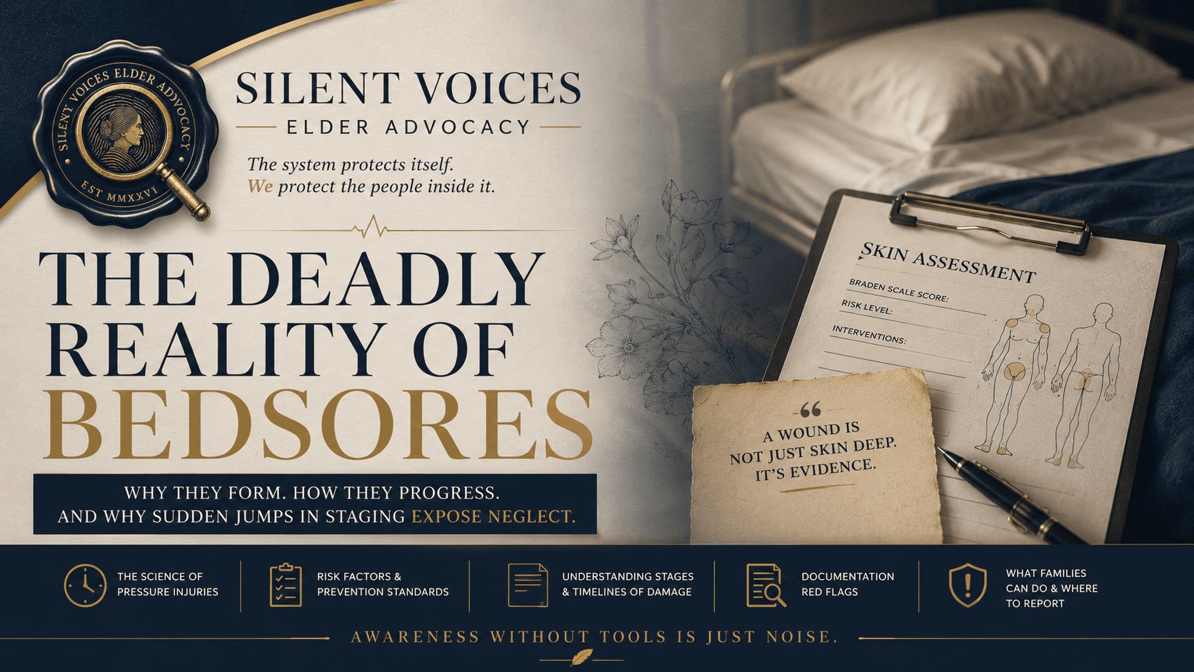
Every year, an estimated 60,000 people in the United States die from complications tied to pressure ulcers [AHRQ]. Among nursing home residents who develop severe infected wounds, mortality rates in clinical literature reach as high as 75 percent in the most serious cases. The Medicare system spends more than two billion dollars annually treating these injuries, though more recent economic analyses place that figure substantially higher [AHRQ]. The most important thing to understand about those numbers: the overwhelming majority of these cases were preventable.
Pressure ulcers, commonly called bedsores, are not a normal part of aging. They are not an inevitable outcome of illness or immobility. When a nursing home resident develops a severe bedsore, particularly one that appears to have progressed rapidly, it is one of the most visible signs of neglect available in elder care.
This article explains why pressure ulcers form, how they progress through the body, what the staging system tells you about a wound’s history, and why a sudden jump in a bedsore’s stage is one of the strongest forensic warning signs that a resident was abandoned by the people paid to care for them.
Why Bedsores Form
At the biological level, a pressure ulcer is an ischemic injury. The word ischemic means the tissue was cut off from its blood supply.
When a person sits or lies in one position without moving, the weight of the body presses down against the mattress or chair. The spots where the body’s bones sit closest to the skin, the tailbone, heels, hips, shoulder blades, and elbows, bear the most pressure. That pressure compresses the tiny blood vessels in the skin. When blood stops flowing, the tissue stops receiving oxygen. If the pressure goes unrelieved, the tissue begins to die [NPIAP].
Pressure ulcers develop from the inside out. Muscle and deep tissue are more sensitive to oxygen deprivation than skin. The damage often starts in the deepest layers and works its way to the surface. This is why some wounds appear to explode over 24 to 48 hours. The visible eruption was not the beginning. It was the end of a process that had already been happening beneath the surface for days.
The body compounds this problem in two ways. First, when tissue is finally reperfused after a long period of oxygen starvation, the sudden return of blood triggers an inflammatory reaction that causes additional cellular damage. Second, frail or malnourished residents have less physiological reserve, meaning their tissues tolerate ischemia for a shorter period before breaking down.
The Forces That Destroy Tissue
Three distinct mechanical forces drive pressure ulcer development. Understanding all three matters because each one tells a different story about what was happening to the resident.
The primary driver. When external force on the skin exceeds what the blood vessels withstand, circulation stops. This is the force controlled by repositioning schedules, specialty mattresses, and pressure-redistributing cushions.
Occurs when skin drags across a surface, most often bed linens. Improper transfers, where a patient is dragged rather than lifted, strip the outer layer of skin. Friction alone does not cause deep wounds, but it removes the skin’s natural defense against everything else.
The most destructive and hardest to see. When the skeleton moves in one direction while the skin stays anchored to the surface, the blood vessels connecting deep tissue to the surface tear from the inside. The skin overhead looks fine. The tissue underneath is already dying. Wounds driven by shear develop deep tunneling beneath the surface, creating hidden cavities of dead tissue that serve as reservoirs for infection [NPIAP].
Moisture accelerates breakdown on top of all three forces. Residents left in soiled briefs for full shifts are exposed to chemical irritants from urine and feces directly against already-compromised skin. Malnutrition and dehydration reduce the body’s capacity to repair even minor damage, turning small injuries into major wounds faster than most families realize.
Who Is Most at Risk
Long-term care facilities are required to evaluate every resident’s risk for pressure ulcers using a standardized clinical tool called the Braden Scale [Braden Scale]. The Braden Scale scores residents across six categories: sensory perception, moisture, activity level, mobility, nutritional intake, and friction and shear exposure. Scores range from 6 to 23. The lower the score, the higher the risk.
A resident scoring 10 or below is classified as high to severe risk, and that score triggers a mandatory clinical response. Facilities are required to order specialized mattresses, establish a documented repositioning schedule tailored to the resident’s individual needs, and manage incontinence aggressively.
| Braden Score | Risk Level | Required Response |
|---|---|---|
| 19 to 23 | No Risk | Routine monitoring, standard skin assessments |
| 15 to 18 | Mild Risk | Preventative barrier creams, weight-shift encouragement |
| 13 to 14 | Moderate Risk | Staff-assisted repositioning schedule, specialty cushions |
| 10 to 12 | High Risk | Mandatory repositioning schedule, alternating air mattress, hourly incontinence management |
| 9 or Below | Severe Risk | Intensive interventions required immediately, specialized support surfaces, meticulous nutrition and hygiene monitoring |
In neglect cases, the Braden Scale becomes one of the most important documents in the medical chart. When a facility correctly scores a resident as high-risk but fails to implement the required interventions, any bedsore that develops is classified by CMS as avoidable [CMS F686]. That classification carries serious regulatory and legal consequences.
The complete absence of Braden assessments in a chart is equally significant. It means the facility never evaluated the resident’s vulnerability at all.
How Bedsores Progress: The Staging System
The National Pressure Injury Advisory Panel classifies pressure ulcers by the depth of tissue destruction. Each stage represents a specific anatomical level of damage.
Skin is intact but red. The redness does not fade when pressed. This signals that blood flow is already compromised. The tissue still has the capacity to recover if pressure is removed immediately.
The outer skin layer breaks down, exposing the dermis. The wound looks like a shallow pink open sore or a fluid-filled blister. It is painful. The damage is still confined to the upper skin layers.
The wound extends through the skin into the subcutaneous fat layer. It looks like a deep crater. Fat tissue is visible. Bone and muscle are not yet exposed, but the wound is serious, prone to infection, and difficult to treat.
Muscle, tendon, bone, or cartilage is exposed or palpable inside the wound. Classified as a Serious Reportable Event [NQF]. Extremely high risk of osteomyelitis and systemic sepsis.
Unstageable: The wound bed is covered by dead tissue, making it impossible to determine true depth without surgical debridement. The wound could be Stage 3 or Stage 4 beneath the surface.
Deep Tissue Pressure Injury (DTPI): Appears as dark purple or maroon discoloration on skin that looks intact. Severe damage is already occurring beneath the surface. DTPIs regularly evolve into Stage 3 or Stage 4 wounds with little warning.
[Infographic Placeholder]
Your chart or visualization embed will go here.
How Fast Do Pressure Ulcers Develop?
One of the biggest misconceptions families hold is that pressure ulcers always develop slowly. The reality is more complicated.
The earliest skin changes, Stage 1 redness, appear within two hours of sustained pressure in a vulnerable resident. A deep tissue pressure injury may develop beneath intact skin during that same window because muscle requires more oxygen than skin and fails first [NPIAP].
Once deep tissue damage has occurred, the wound often becomes visible over the next 24 to 72 hours. What looks like a sudden Stage 3 or Stage 4 ulcer frequently represents tissue that had already been dying beneath the surface for several days.
When a nurse documents a Stage 3 wound for the first time, the chart is recording when the injury was discovered, not when it developed. The biological injury began earlier. One of the key questions in any investigation is whether staff missed earlier warning signs or failed to examine the resident’s skin altogether. “It wasn’t there yesterday” is not always medically accurate, and it is not a defense.
The Sudden Jump Is a Forensic Warning
The staging system moves in one direction. A wound does not skip biology.
Pressure ulcers do not materialize overnight as massive Stage 4 craters. The biology of tissue destruction follows a sequence: capillary compromise, then superficial breakdown, then fat layer necrosis, then bone and muscle exposure. That progression takes days to weeks of sustained, unrelieved pressure.
When a nursing home’s medical record shows a patient with intact skin on Monday and a foul-smelling Stage 3 or Stage 4 wound by Friday, one of two things happened. Either the patient was left immobile for extended periods without anyone repositioning or assessing them, or the documentation in the chart was fabricated.
A sudden jump in staging is not a medical mystery. It is a gap in care that the documentation was either not capturing or was actively concealing. Forensic nurses and plaintiff attorneys treat these jumps as the opening of an investigation, not the conclusion of one.
How Records Get Falsified
Repositioning logs, called turning sheets, require nursing assistants to initial each cycle as proof the resident was moved. In understaffed facilities, these logs are frequently completed at the end of a shift, in bulk, for care that was never provided. Wound care professionals and legal nurses call this pencil-whipping. Three patterns show up repeatedly in neglect cases.
Identical initials recorded at precise intervals across a twelve-hour period are a red flag. So are entries showing a patient repositioned at hours when transport records confirm the patient was off-site at a dialysis appointment.
A sudden Stage 4 presentation with no documented Stage 1 or Stage 2 wound preceding it means the earlier warning signs were either never observed or deliberately ignored.
A wound charted at the same dimensions week after week while the resident’s condition deteriorates is documentation designed to avoid triggering wound care protocols and family notifications the facility was legally required to provide [CMS F686]. When a hospital admission finally occurs due to sepsis, the independent physician finds a wound completely inconsistent with the nursing home’s charted history.
The “Unavoidable” Defense and Its Limits
Facilities sometimes invoke a clinical concept called the Kennedy Terminal Ulcer when defending pressure injury claims. While terminal skin failure is recognized in medicine, the diagnosis remains controversial, and experts evaluate each case individually using the resident’s overall clinical picture, wound location, timing, and documented preventive care [NPIAP Position Statements].
The physiological question at the center of every KTU defense is straightforward. Terminal decline is a systemic condition. If a dying patient’s skin truly fails because of global circulatory collapse, the breakdown would appear across all bony prominences simultaneously. Every pressure point would show signs of failure.
When a patient dies with a single, massive sacral ulcer and their heels, hips, and shoulder blades are entirely intact, the wound location tells a specific story. Pressure was concentrated on the sacrum. The “unavoidable terminal ulcer” defense struggles when only one location broke down, because uniform physiological dying does not produce a single localized wound.
From Wound to Death: The Sepsis Pathway
A Stage 3 or Stage 4 wound left untreated becomes a clinical emergency on a predictable timeline [CDC Sepsis Data]. The open wound fills with necrotic tissue. The surrounding tissue develops cellulitis. As the infection deepens, it reaches exposed bone, causing osteomyelitis.
Among nursing home residents who die from pressure ulcer complications, clinical literature documents that 50 percent succumb within six weeks of the wound’s first appearance. This is not a rare complication of advanced illness. It is a predictable outcome of abandonment.
What Families Can Do Now
If your family member lives in a nursing home, these are the specific actions you take now.
Federal regulations require these records to exist [CMS Residents’ Rights]. If a facility refuses to produce them or they are incomplete, document that refusal in writing.
If staff cannot tell you your family member’s score, ask for the last documented assessment date. If none exists, file a complaint.
Photograph any redness, skin tears, or open wounds. Date and time-stamp your photos. This creates an independent record the facility cannot retroactively alter.
Ask whether your family member is on a pressure-redistributing mattress. Two hours has long served as the traditional clinical standard for repositioning immobile residents, though some patients require more frequent repositioning based on individual risk factors and their care plan [NPIAP]. What matters is that the facility has a documented, individualized schedule and that staff are following it.
Reporting in Arkansas
If you suspect neglect related to a pressure ulcer in Arkansas, these are the agencies with authority to investigate. Photograph the wound before you leave the building. Request a complete copy of the medical chart, including repositioning logs and care plans, before alerting the facility you intend to file a complaint. Once they know, records are at risk.
| Agency | Scope | Contact |
|---|---|---|
| Adult Protective Services Arkansas DHS |
Maltreatment of adults 18 and older living in the community | 1-800-482-8049 |
| Office of Long Term Care Arkansas DHS |
Complaints and F686 violations for residents in licensed long-term care facilities | 1-800-582-4887 complaints.OLTC@arkansas.gov |
| Long-Term Care Ombudsman Arkansas Ombudsman Program |
Independent resident rights advocate, complaint investigation, Nursing Home Reform Act education | 1-501-682-8952 |
A pressure ulcer is not simply a wound. It is a timeline written on the body. When the medical record tells one story and the anatomy tells another, the body is usually the more reliable witness.
Stage 4 pressure ulcers are classified as never events [NQF] for a reason. You have the right to request records, file a complaint, and demand an independent medical evaluation. The staging system exists to measure what happened. Use it.
Nathalie Frias is a certified electronic health records specialist and founder of Silent Voices Elder Advocacy, a platform for elder care accountability and fiction.
Her writing translates clinical documentation into evidence families and advocates can use to expose neglect, financial exploitation, and regulatory failures in long-term care.
Learn more at nathaliefrias.netGet elder care accountability resources, new investigations, and practical tools sent directly to you.
Subscribe to Silent Voices




Leave a Reply